Over recent years, cases of the ‘flesh-eating’ Buruli ulcer have increased significantly in Victoria and Far North Queensland (NSW Health 2025).
Prior to 2015, Victoria recorded fewer than 100 cases of Buruli ulcer each year. However, since 2017, the number of annual cases has consistently ranged between 200 and 370 (Muhi et al. 2025).
But what exactly is this illness, why is it described as ‘flesh-eating’ and is it a cause for concern?
What is Buruli Ulcer?

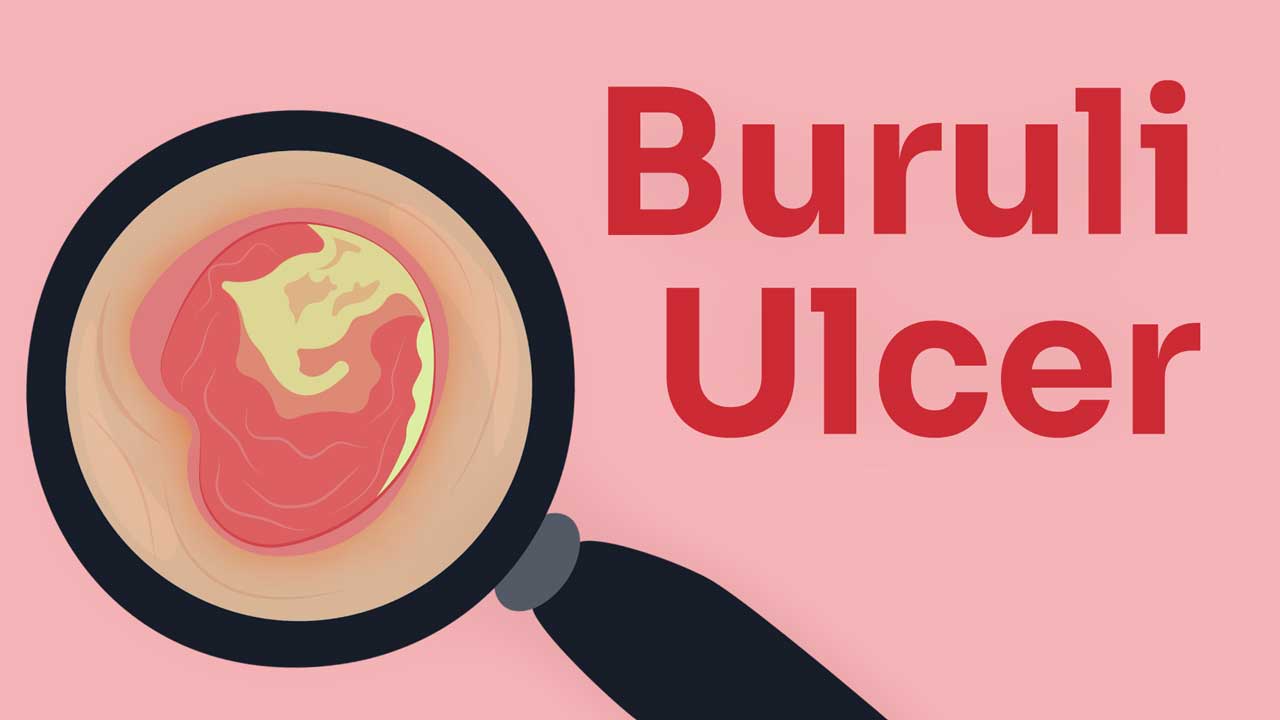
Buruli ulcer is a bacterial infection of the skin and soft tissue that causes progressive, non-healing wounds (Wong et al. 2024).
Buruli ulcer is caused by the bacteria Mycobacterium ulcerans, which belongs to the same family as the bacteria responsible for leprosy and tuberculosis (Healthdirect 2023).
Mycobacterium ulcerans bacteria produce a toxin known as mycolactone that weakens the body’s immune response and is toxic to the cells, resulting in extensive tissue damage, ulceration and skin loss. If left untreated, this ulceration can extend to the nerves, blood vessels, muscles and even bone (Better Health Channel 2025; Brown & Altman 2021).
For this reason, the condition is often described as a ‘flesh-eating ulcer’ (Healthdirect 2023).
Buruli ulcer has been detected in 33 countries. Most cases in Australia occur in Victoria, Queensland and the Northern Territory (Better Health Channel 2025).
What Causes Buruli Ulcer?
Mycobacterium ulcerans bacteria are found naturally in the environment and have been detected in mosquitoes, possum faeces, vegetation and areas of swampy and stagnant water (Better Health Channel 2025; Healthdirect 2023).
It’s currently not fully understood how humans become infected with Mycobacterium ulcerans. However, most cases occur in areas where mosquitoes and possums have been found to be carrying the bacteria (Better Health Channel 2025; VIC DoH 2024).
It’s speculated that possums act as a reservoir for Mycobacterium ulcerans, with mosquitoes transmitting the bacteria to humans through bites (Brown & Altman 2021).
The bacteria can’t be transmitted from person to person (Healthdirect 2023).
Symptoms of Buruli Ulcer
Symptoms typically develop between one to nine months after contracting the infection (Muhi et al. 2025).
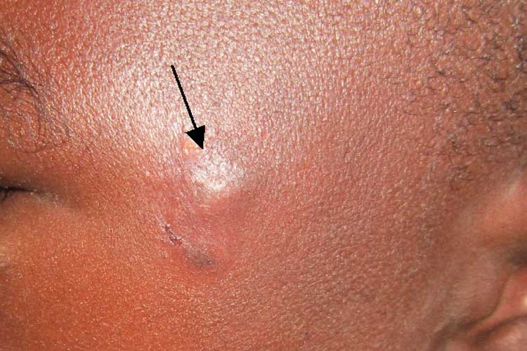
The first sign of the infection is a firm, painless nodule on the skin. The nodule will most commonly appear on the arms, legs or face. It will typically be between 1 and 2 cm in diameter, resembling a mosquito or spider bite (Healthdirect 2023; Brown & Altman 2021; Better Health Channel 2025).

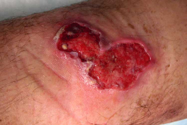
Over the next days or weeks, the nodule will gradually grow larger and may form a crusty, non-healing scab that eventually ulcerates (Better Health Channel 2025).
The resulting ulcer is usually painless with undermined edges, signifying tissue damage under the skin. If left untreated, the ulcer will continue to extend and may destroy other tissue, muscle and bone (Healthdirect 2023; Brown & Altman 2021).
Occasionally, the infection will present without ulceration. The patient may experience localised pain and swelling accompanied by a fever, raised lumps, or thickened or raised areas of flat skin (Better Health Channel 2025).
Complications of Buruli Ulcer
- Significant tissue destruction (up to 15% of the skin surface)
- Secondary infection
- Osteomyelitis (bone infection)
- Metastatic lesions (spread of wounds to different parts of the body)
- Scarring
- Secondary lymphoedema due to fluid retention
- Contractures.
(Brown & Altman 2021)
Lifelong disability, disfigurement and socioeconomic burden are common consequences of the infection, particularly if recognition is slow and access to treatment is limited (WHO 2023; Wong et al. 2024).
Diagnosis of Buruli Ulcer
The fastest, easiest and most accurate way to diagnose Buruli ulcer is via polymerase chain reaction (PCR) test to detect the presence Mycobacterium ulcerans, using a swab or tissue biopsy of the affected area (O'Brien et al. 2019; Healthdirect 2023).
The characteristic ulcer might resemble lesions or wounds caused by diabetes, or arterial or venous insufficiency. It’s important to rule out these differential diagnoses during assessment (WHO 2023).
Early diagnosis is crucial in ensuring prompt treatment and preventing the progression of the illness (Healthdirect 2023).
Treatment of Buruli Ulcer
Treatment comprises both antibiotics and complementary management (WHO 2023).
Currently, the recommended course of antibiotics is a combination of rifampicin and clarithromycin (WHO 2023).
An alternative combination of rifampicin and moxifloxacin is commonly used in Australia, but more research is needed on its efficacy (WHO 2023).
Complementary treatments may involve:
- Wound and lymphoedema management
- Surgical intervention (debridement and skin grafting) to speed up the healing process
- In severe cases, physiotherapy to prevent disability
- Long-term rehabilitation (in cases of disability).
(WHO 2023)
Preventing Buruli Ulcer

Due to the mode of transmission being unknown, it’s not understood how exactly Buruli ulcer can be prevented. Despite this, protecting against potential sources of infection, such as soil and mosquito bites, may help to reduce the risk (Better Health Channel 2025). Precautions may include:
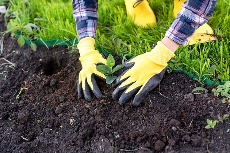
- Wearing gloves, long-sleeved shirts and long pants when working outdoors
- Using insect repellents
- Using plasters to protect cuts and abrasions
- Washing and covering any wounds sustained while working outdoors
- Seeking medical advice in the case of a slow-healing skin lesion.
(Better Health Channel 2025)
Overall, however, the risk of Buruli infection in Australia is low (Better Health Channel 2025).
Test Your Knowledge
Question 1 of 3
What kind of infection is Buruli ulcer?
Topics
References
- Better Health Channel 2025, Buruli Ulcer, Victoria State Government, viewed 25 July 2025, https://www.betterhealth.vic.gov.au/health/healthyliving/Buruli-ulcer
- Brown, SC & Altman, K 2021, Buruli Ulcer, Medscape, viewed 25 July 2025, https://emedicine.medscape.com/article/1104891-overview#a4
- Healthdirect 2023, Buruli Ulcer, Australian Government, viewed 25 July 2025, https://www.healthdirect.gov.au/buruli-ulcer
- Johnson, PDR 2019, ‘Buruli Ulcer in Australia’, in G Pluschke & K Röltgen (eds.) Buruli Ulcer: Mycobacterium Ulcerans Disease, viewed 24 July 2025, https://www.ncbi.nlm.nih.gov/books/NBK553830/
- Muhi, S, Cox, VRV, O'Brien, M et al. 2025, ‘Management of Mycobacterium ulcerans Infection (Buruli Ulcer) in Australia: Consensus Statement’, Med J Aust., viewed 24 July 2025, https://www.mja.com.au/journal/2025/222/11/management-mycobacterium-ulcerans-infection-buruli-ulcer-australia-consensus
- NSW Health 2025, Buruli Ulcer, New South Wales Government, viewed 24 July 2025, https://www.health.nsw.gov.au/Infectious/factsheets/Pages/buruli-ulcer.aspx
- O'Brien, DP, Globan, M, Fyfe, JM et al. 2019, ‘Diagnosis of Mycobacterium Ulcerans Disease: Be Alert to the Possibility of Negative Initial PCR Results’, Med J Aust., vol. 210, no. 9, viewed 25 July 2025, https://www.mja.com.au/journal/2019/210/9/diagnosis-mycobacterium-ulcerans-disease-be-alert-possibility-negative-initial
- Victoria Department of Health 2024, Beating Buruli in Victoria, Victoria State Government, viewed 25 July 2025, https://www.health.vic.gov.au/infectious-diseases/beating-buruli-in-victoria
- World Health Organisation 2023, Buruli Ulcer (Mycobacterium Ulcerans Infection), WHO, viewed 25 July 2025, https://www.who.int/news-room/fact-sheets/detail/buruli-ulcer-(mycobacterium-ulcerans-infection)
 New
New 
